Histopathological Changes in Diabetic Pancreas
Aysenur Kaya (Author), Murat Cetin Ragbetli (Author)
Release Date: 2023-09-14
Histopathological changes in the diabetic pancreas are characterized by several key alterations that impact its structure and function. In type 1 diabetes mellitus (T1DM), autoimmune destruction of insulin-producing beta cells within the pancreatic islets results in their selective loss, termed insulitis. This process involves infiltration of immune cells such as T lymphocytes and macrophages into [...]
Media Type
PDF
Buy from
Price may vary by retailers
| Work Type | Book Chapter |
|---|---|
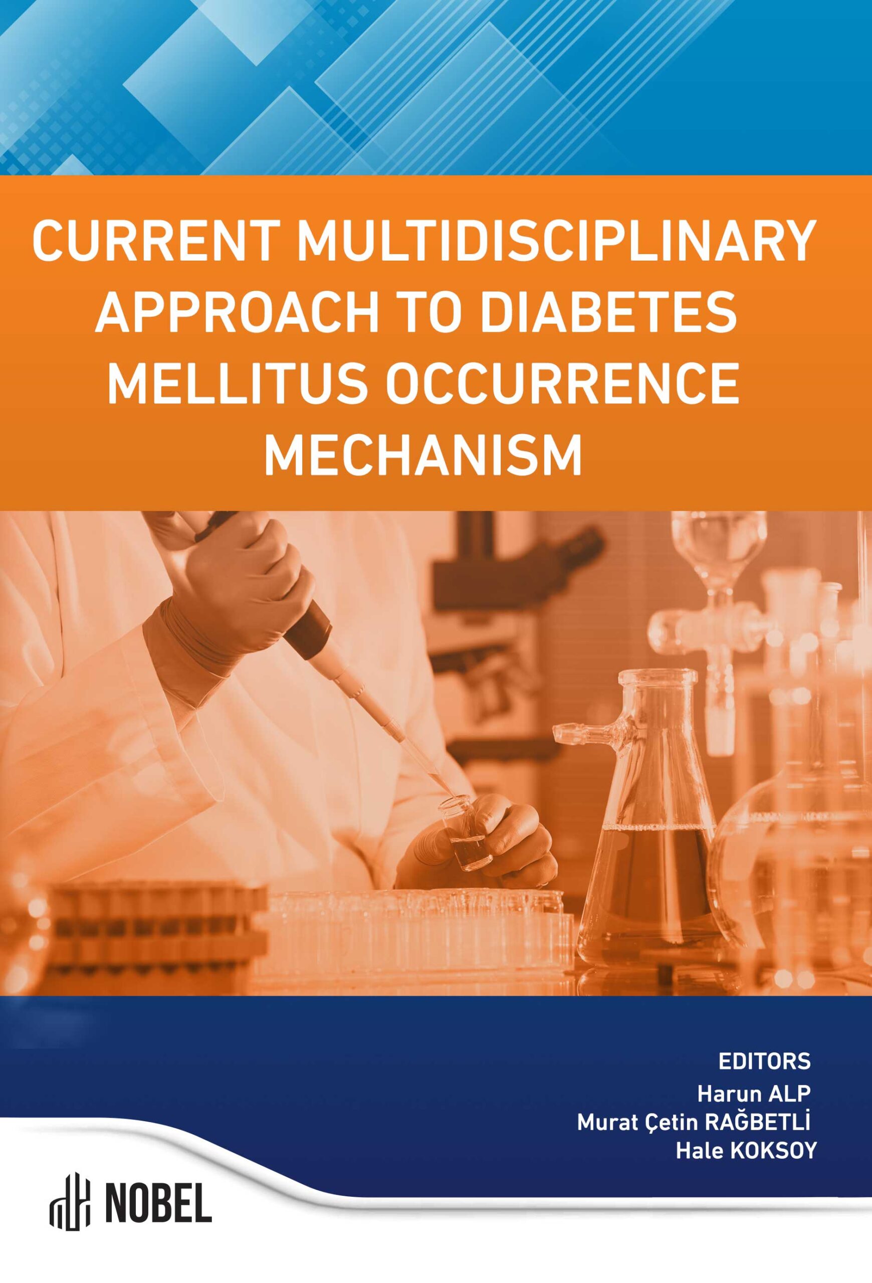
| Published in | Current Multidisciplinary Approach to Diabetes Mellitus Occurrence Mechanism |
| First Page | 29 |
| Last Page | 36 |
| DOI | https://doi.org/10.69860/nobel.9786053359104.3 |
| ISBN | 978-605-335-910-4 (PDF) |
| Language | ENG |
| Page Count | 8 |
| Copyright Holder | Nobel Tıp Kitabevleri |
| License | https://nobelpub.com/publish-with-us/copyright-and-licensing |
Aysenur Kaya (Author)
Research Assistant, Karamanoglu Mehmetbey University
https://orcid.org/0000-0002-3878-0087
Murat Cetin Ragbetli (Author)
Professor, Karamanoglu Mehmetbey University
https://orcid.org/0000-0002-8189-264X
Richardson SJ, Pugliese A. 100 YEARS OF INSULIN: Pancreas pathology in type 1 diabetes: an evolving story. J Endocrinol. 2021;252(2):R41-r57.
Huang YH, Sun MJ, Jiang M, Fu BY. Immunohistochemical localization of glucagon and pancreatic polypeptideon rat endocrine pancreas: coexistence in rat islet cells. Eur J Histochem. 2009;53(2):81-5.
Sakata N, Yoshimatsu G, Kodama S. Development and Characteristics of Pancreatic Epsilon Cells. Int J Mol Sci. 2019;20(8).
Leung PS. Overview of the pancreas. Adv Exp Med Biol. 2010;690:3-12.
Henry BM, Skinningsrud B, Saganiak K, Pękala PA, Walocha JA, Tomaszewski KA. Development of the human pancreas and its vasculature - An integrated review covering anatomical, embryological, histological, and molecular aspects. Ann Anat. 2019;221:115-24.
Bertelli E, Bendayan M. Association between endocrine pancreas and ductal system. More than an epiphenomenonof endocrine differentiation and development? J Histochem Cytochem. 2005;53(9):1071-86.
Bensley RR. Studies on the pancreas of the guinea pig. Am J Anat. 1911;12(3):297-388.
van Suylichem PT, Wolters GH, van Schilfgaarde R. Peri-insular presence of collagenase during islet isolation procedures. J Surg Res. 1992;53(5):502-9.
Suzuki T, Kadoya Y, Sato Y, Handa K, Takahashi T, Kakita A, et al. The expression of pancreatic endocrine markers in centroacinar cells of the normal and regenerating rat pancreas: their possible transformation to endocrine cells. Arch Histol Cytol. 2003;66(4):347-58.
Kulkarni RN. The islet beta-cell. Int J Biochem Cell Biol. 2004;36(3):365-71.
Erejuwa OO, Sulaiman SA, Wahab MS, Salam SK, Salleh MS, Gurtu S. Antioxidant protective effect of glibenclamide and metformin in combination with honey in pancreas of streptozotocin-induced diabetic rats. Int J Mol Sci. 2010;11(5):2056-66.
Busnardo AC, DiDio LJ, Tidrick RT, Thomford NR. History of the pancreas. Am J Surg. 1983;146(5):539-50.
Marshall SM. The pancreas in health and in diabetes. Diabetologia. 2020;63(10):1962-5.
Williams AJ, Thrower SL, Sequeiros IM, Ward A, Bickerton AS, Triay JM, et al. Pancreatic volume is reduced in adult patients with recently diagnosed type 1 diabetes. J Clin Endocrinol Metab. 2012;97(11):E2109-13.
Damond N, Engler S, Zanotelli VRT, Schapiro D, Wasserfall CH, Kusmartseva I, et al. A Map of Human Type 1 Diabetes Progression by Imaging Mass Cytometry. Cell Metab. 2019;29(3):755-68.e5.
Shi YC, Pan TM. Antioxidant and pancreas-protective effect of red mold fermented products on streptozotocin-induced diabetic rats. J Sci Food Agric. 2010;90(14):2519-25.
Davison LJ. Diabetes mellitus and pancreatitis--cause or effect? J Small Anim Pract. 2015;56(1):50-9.
In’t Veld P. Insulitis in human type 1 diabetes: The quest for an elusive lesion. Islets. 2011;3(4):131-8.
Nigi L, Maccora C, Dotta F, Sebastiani G. From immunohistological to anatomical alterations of human pancreas in type 1 diabetes: New concepts on the stage. Diabetes Metab Res Rev. 2020;36(4):e3264.
Yagihashi S. Diabetes and pancreas size, does it matter? J Diabetes Investig. 2017;8(4):413-5.
Al-Mrabeh A, Hollingsworth KG, Steven S, Taylor R. Morphology of the pancreas in type 2 diabetes: effectof weight loss with or without normalisation of insulin secretory capacity. Diabetologia. 2016;59(8):1753-9.
Gastaldelli A, Cusi K, Pettiti M, Hardies J, Miyazaki Y, Berria R, et al. Relationship between hepatic/ visceral fat and hepatic insulin resistance in nondiabetic and type 2 diabetic subjects. Gastroenterology. 2007;133(2):496-506.
Maša Skelin K, Jurij D, Ismael V-A, Andraž S, Saška L. Application of Transmission Electron Microscopy to Detect Changes in Pancreas Physiology. In: Mohsen M, editor. Electron Microscopy. Rijeka: IntechOpen; 2022. p. Ch. 7.
Petrov MS, Taylor R. Intra-pancreatic fat deposition: bringing hidden fat to the fore. Nat Rev Gastroenterol Hepatol. 2022;19(3):153-68.
Abdul-Hamid M, Moustafa N. Protective effect of curcumin on histopathology and ultrastructure of pancreasin the alloxan treated rats for induction of diabetes. J Basic Appl Zool. 2013;66(4):169-79.
Halliwell B, Chirico S. Lipid peroxidation: its mechanism, measurement, and signifi cance. Am J Clin Nutr. 1993;57(5 Suppl):715S-24S; discussion 24S-25S.
Attia AA. Histological and electron microscopic studies of the effect of beta-carotene on the pancreas ofstreptozotocin (STZ)-induced diabetic rats. Pak J Biol Sci. 2009;12(4):301-14
| onix_3.0::thoth | Thoth ONIX 3.0 |
|---|---|
| onix_3.0::project_muse | Project MUSE ONIX 3.0 |
| onix_3.0::oapen | OAPEN ONIX 3.0 |
| onix_3.0::jstor | JSTOR ONIX 3.0 |
| onix_3.0::google_books | Google Books ONIX 3.0 |
| onix_3.0::overdrive | OverDrive ONIX 3.0 |
| onix_2.1::ebsco_host | EBSCO Host ONIX 2.1 |
| csv::thoth | Thoth CSV |
| json::thoth | Thoth JSON |
| kbart::oclc | OCLC KBART |
| bibtex::thoth | Thoth BibTeX |
| doideposit::crossref | CrossRef DOI deposit |
| onix_2.1::proquest_ebrary | ProQuest Ebrary ONIX 2.1 |
| marc21record::thoth | Thoth MARC 21 Record |
| marc21markup::thoth | Thoth MARC 21 Markup |
| marc21xml::thoth | Thoth MARC 21 XML |

