Craniosynostosis: Clinical Characteristics, Molecular Mechanisms and Treatment
Suray Pehlivanoglu (Author), Sebnem Pehlivanoglu (Author)
Release Date:
Craniosynostosis is a congenital condition marked by the early fusion of one or more cranial sutures. Cranial sutures are fibrous tissues that connect the skull bones. They play a crucial role in ensuring bone formation at the edges of the calvarial bones, which move apart to facilitate the passage of the head through the birth [...]
Media Type
Buy from
Price may vary by retailers
| Work Type | Book Chapter |
|---|---|
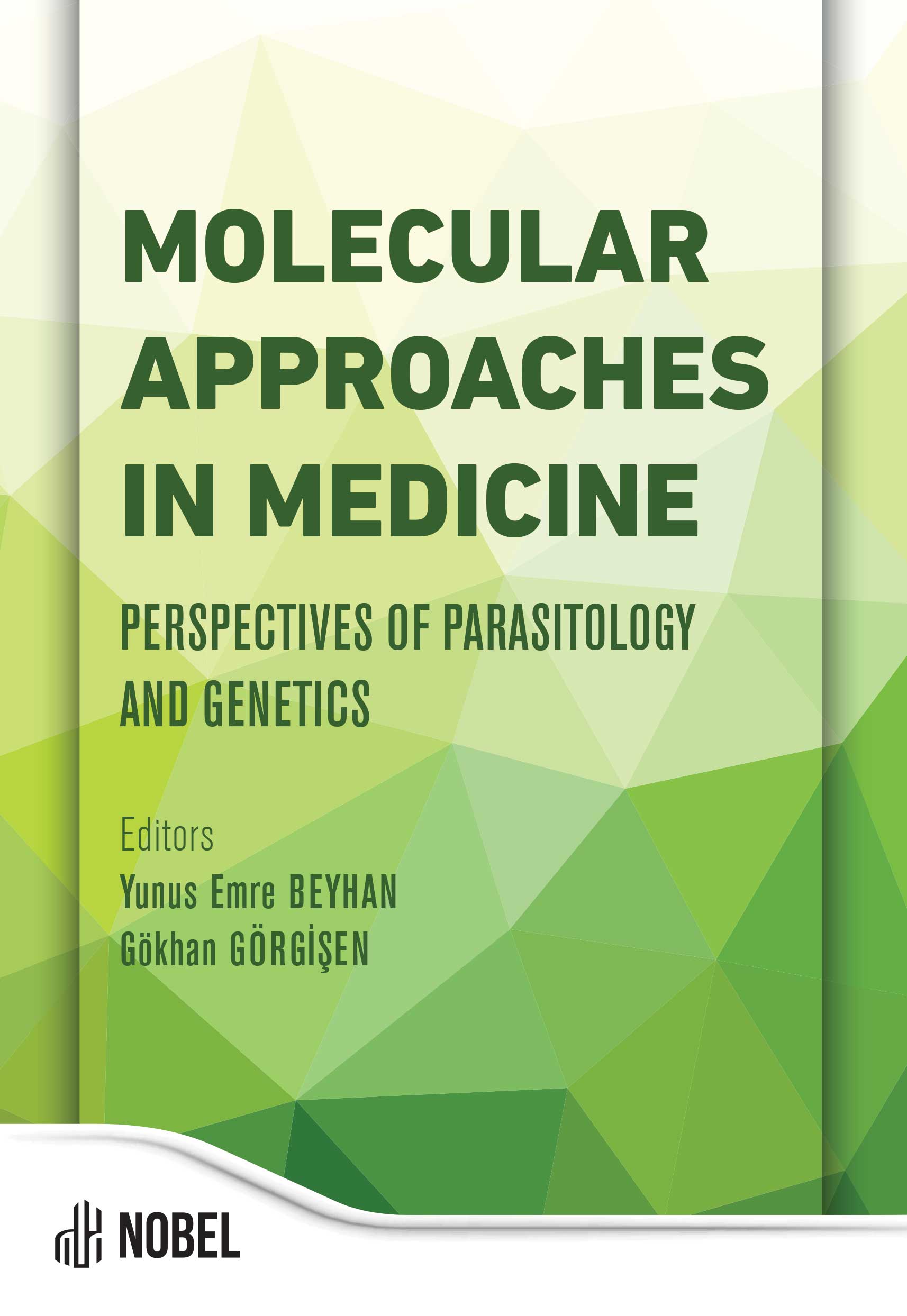
| Published in | Molecular Approaches in Medicine |
| First Page | 109 |
| Last Page | 133 |
| DOI | https://doi.org/10.69860/nobel.9786053359524.6 |
| Page Count | 25 |
| Copyright Holder | Nobel Tıp Kitabevleri |
| License | https://nobelpub.com/publish-with-us/copyright-and-licensing |
Suray Pehlivanoglu (Author)
Associate Professor, Necmettin Erbakan University
https://orcid.org/0000-0001-7422-2974
3Assoc. Prof. Dr. Suray Pehlivanoglu is a dedicated scientist with international publications, specializing in congenital diseases, cellular therapies, and cancer biology. In the field of congenital diseases, he has contributed to the establishment of the craniosynostosis mutation spectrum in Turkish cases. Additionally, he is conducting research on alternative signaling pathways leading to congenital primary immunodeficiencies, supported by the Turkish Health Institutes Presidency. The TUBITAK project he worked on regarding induced pluripotent stem cells and cellular therapies has been honored with the Süreyya Tahsin Aygün Award under the auspices of the Turkish Academy of Sciences. In the realm of cancer biology, he investigates cell migration, fibrosis, and associated signaling pathways in the context of the epithelial-mesenchymal transition process. As a result of his studies on the discovery of anticancer and antimetastatic agents, his identified agents have been patented by the Turkish Patent Institute, and international patent processes are ongoing.
Sebnem Pehlivanoglu (Author)
Necmettin Erbakan University
https://orcid.org/0000-0003-0817-0891
Anderson, P.J., Cox, T.C., Smithers, L., et al. (2007). Somatic FGFR and TWIST Mutations are not a Common Cause of Isolated Nonsyndromic Single Suture Craniosynostosis. The Journal of Craniofacial Surgery.,
18:312- 314.
Barkovich, M.J., Xu, D., Desikan, R.S., et al. (2018). Pediatric neuro MRI: tricks to minimize sedation. Pediatr Radio.,l 48, 50-55.
Barone, C.M., Jimenez, D.F. (1999). Endoscopic craniectomy for early correction of craniosynostosis. Plast Reconstr Surg.,104:1965-1973, discussion 1974-1975.
Beck, J., Parent, A., Angel, M.F. (2002). Chronic headache as a sequela of rigid fixation for craniosynostosis. J Craniofac Surg.,13: 327-330.
Boyadjiev, S.A. (2007). Genetic analysis of non-syndromic craniosynostosis. Orthod Craniofacial Res., 10:129–137.
Clauser, L., Galiè, M., Hassanipour, A., et al. (2000). Saethre-Chotzen syndrome: review of the literature and report of a case. J Craniofac Surg., Sep;11(5):480-6.
Cohen, M.M. Jr. (2005). Editorial: Perspectives on Craniosynostosis. Am.J. Genet. A. 2005.136(4):313-326.
Cohen, M.M. (1991). Etiopathogenesis of craniosynostosis. Neurosurg Clin. N. Am. 2(3):507-513.
Cohen, M. M., Jr. (2012). No man’s craniosynostosis: the arcana of sutural knowledge. J Craniofac Surg, 23, 338-342.
Cohen, M., Michael and MacLean, Ruth E. (2000). Craniosynostosis: diagnosis, evaluation, and management. New York: Oxford University Press.
Connerneya, J.J., Spicer, D.B. (2011). Signal Transduction Pathways and Their Impairment in Syndromic Craniosynostosis. Basel: Karger.
Coutu, D. L., Galipeau, J. (2011). Roles of FGF signaling in stem cell self- renewal, senescence and aging. Aging (Albany NY), 3, 920-933.
Crouzon, O. (1912). Dysostose cranio-faciale hereditaire. Bull Mem Soc Med Hop Paris, 33, 545-555.
Cunningham, M. L., Heike, C. L. (2007). Evaluation of the infant with an abnormal skull shape. Curr Opin Pediatr, 19, 645-651.
Çeltikçi, E., Börcek, A. Ö., Baykaner, M. K. (2013). Kraniyosinostozlar. Türk Nöroşirürji Dergisi, 23, 132-137.
David, M., Ornitz Marie J.P. (2002). FGF signaling pathways in endochondral and intramembranous bone development and human genetic disease. Genes & Dev., 16:1446-1465.
Dienstmann, R., Rodon, J., Prat, A., et. al. (2014). Genomic aberrations in the FGFR pathway: opportunities for targeted therapies in solid tumors. Ann Oncol, 25, 552-563.
Dubois, J., Garel, L. (1999). Imaging and therapeutic approach of hemangiomas and vascular malformations in the pediatric age group. Pediatr Radiol. 1999;29(12):879-893.
Ertosun, M.G., Pehlivanoglu, S., Dilmac, S., Tanriover, G., Ozes, O.N. (2020). AKT-mediated phosphorylation of TWIST1 is essential for breast cancer cell metastasis. Turk J Biol. 19;44(4):158-165.
Guillemot, F., Zimmer, C. (2011). From cradle to grave: the multiple roles of fibroblast growth factors in neural development. Neuron, 71, 574- 588.
Hatch, N. E., Hudson, M., Seto, M. L., et al. (2006). Intracellular retention, degradation, and signaling of glycosylation-deficient FGFR2 and craniosynostosis syndrome-associated FGFR2C278F. J Biol Chem, 281, 27292-27305.
Hehr, U., Muenke, M. (1999). Craniosynostosis Syndromes: From Genes to Premature Fusion of Skull Bones. Mol. Genet. and Met., 68:139–151.
Heuze, Y., Holmes, G., Peter, I. et. al. (2014). Closing the Gap: Genetic and Genomic Continuum from Syndromic to Nonsyndromic Craniosynostoses. Curr Genet Med Rep, 2, 135-145.
http://www.ensembl.org/Homo_sapiens/.
Huang, N., Pandey, A. V., Agrawal, V., et. al. (2005). Diversity and function of mutations in p450 oxidoreductase in patients with Antley-Bixler syndrome and disordered steroidogenesis. Am J Hum Genet, 76, 729-749.
Jay, S., Wiberg, A., Swan, M., et. al. (2013). The fibroblast growth factor receptor 2 p.Ala172Phe mutation in Pfeiffer syndrome--history repeating itself. Am J Med Genet A, 161A, 1158-1163.
Jenkins, D., Seelow, D., Jehee, F. S., et. al. (2007). RAB23 mutations in Carpenter syndrome imply an unexpected role for hedgehog signaling in cranial-suture development and obesity. Am J Hum Genet, 80, 1162-1170.
Johnson, D., Wilkie, A. O. (2011). Craniosynostosis. Eur J Hum Genet, 19, 369-376.
Kabbani H, Raghuveer TS. (2004). Craniosynostosis. Am Fam Physician. 15;69(12):2863-70.
Kajdic, N., Spazzapan, P., Velnar, T. (2018). Craniosynostosis – Recognition, clinical characteristics, and treatment. Bosn J Basic Med Sci., (2):110-116.
Kan, S. H., Elanko, N., Johnson, D., et. al. (2002). Genomic screening of fibroblast growth-factor receptor 2 reveals a wide spectrum of mutations in patients with syndromic craniosynostosis. Am J Hum Genet, 70, 472-486.
Kane, A.A. (2004). An Overview of Craniosynostosis. JPO Journal of Prosthetics & Orthotics, 16, 50-55.
Khanna, P. C., Thapa, M. M., Iyer, R. S., et. al. (2011). Pictorial essay: The many faces of craniosynostosis. Indian J Radiol Imaging., 21, 49-56.
Kimonis, V., Gold, J. A., Hoffman, T. L., et. al. (2007). Genetics of craniosynostosis. Semin Pediatr Neurol., 14, 150-161.
Kotrikova, B., Krempien, R., Freier, K., and Mühling, J. (2007). Diagnostic imaging in the management of craniosynostoses. Eur Radiol., 2007.17:1968–1978.
Kress, W., Schropp, C., Lieb, G., et. al. (2006). Saethre-Chotzen syndrome caused by TWIST 1 gene mutations: functional differentiation from Muenke coronal synostosis syndrome. Eur J Hum Genet., 14, 39-48.
Lajeunie, E., Le Merrer, M., Bonaıti-Pellie, C., Marchac, D., Renier, D. (1986). Genetic study of scaphocephaly. Am J Med Genet., 62:282–285.
Levi, B., Wan, D.C., Wong, V.W., et. al. (2012). Cranial Suture Biology: From Pathways to Patient Care. Journal of Craniofacial Surgery., 23, 13.
Mansukhani, A., Bellosta, P., Sahni, M., et al. (2000). Signaling by fibroblast growth factors (FGF) and fibroblast growth factor receptor 2 (FGFR2)-activating mutations blocks mineralization and induces apoptosis in osteoblasts. J Cell Biol., 149, 1297-1308.
Martinez-Abadias, N., Heuze, Y., Wang, Y., et. al. (2011). FGF/FGFR signaling coordinates skull development by modulating magnitude of morphological integration: evidence from Apert syndrome mouse models. PLoS One, 6, e26425.
Mohammadi, M., Olsen, S. K., Ibrahimi, O. A. (2005). Structural basis for fibroblast growth factor receptor activation. Cytokine Growth Factor Rev., 16, 107-137.
Musolf AM, Justice CM, Erdogan-Yildirim Z, et. al. (2024). Whole genome sequencing identifies associations for nonsyndromic sagittal craniosynostosis with the intergenic region of BMP2 and noncoding RNA gene LINC01428. Sci Rep., Apr 12;14(1):8533.
Nur, B.G, Pehlivanoğlu, S., Mıhçı, E.,et al. (2014). Clinicogenetic study of Turkish patients with syndromic craniosynostosis and literature review. Pediatr Neurol., May;50(5):482-90.
Ornitz, D. M. (2000). FGFs, heparan sulfate and FGFRs: complex interactions essential for development. Bioessays, 22, 108-112.
Park, J., Park, O. J., Yoon, W. J., et. al. (2012). Functional characterization of a novel FGFR2 mutation, E731K, in craniosynostosis. J Cell Biochem., 113, 457-464.
Passos-Bueno, M. R., Serti Eacute, A. E., Jehee, F. S., et. al. (2008). Genetics of craniosynostosis: genes, syndromes, mutations and genotype-phenotype correlations. Front Oral Biol., 12, 107-143.
Patil, S., Biassoni, L., Borgwardt, L. (2007). Nuclear medicine in pediatric neurology and neurosurgery: epilepsy and brain tumors. In Seminars in nuclear medicine, 37, 5, 357-381.
Ramirez-Schrempp, D., Vinci, R.J., Liteplo, A.S. (2011). Bedside ultrasound in the diagnosis of skull fractures in the pediatric emergency department. Pediatr Emerg Care., 27(4):312-314.
Ranger, A., Chaudhary, N., Matic, D. (2010). Craniosynostosis involving the squamous temporal sutures: a rare and possibly underreported etiology for cranial vault asymmetry. J Craniofac Surg, 21, 1547-1550.
Reardon, W., Smith, A., Honour, J. W., et. al. (2000). Evidence for digenic inheritance in some cases of Antley-Bixler syndrome? J Med Genet., 37, 26- 32.
Reardon, W., Winter, R. M., Rutland, P., et. al. (1994). Mutations in the fibroblast growth factor receptor 2 gene cause Crouzon syndrome. Nat Genet, 8, 98-103.
Richman, J.M. (1995). Craniofacial genetics makes headway. Curr. Biol. 5:4.
Roscioli, T., Elakis, G., Cox, T.C., et al. (2013). Genotype and clinical care correlations in craniosynostosis: findings from a cohort of 630 Australian and New Zealand patients. Am J Med Genet C Semin Med Genet., Nov,163C(4):259-70.
Sandhaus, H., Johnson, M.D. (2021) .Distraction osteogenesis in craniosynostosis. Curr Opin Otolaryngol Head Neck Surg., Aug 1;29(4):304-313.
Seto, M.L,. Hing, A.V., Chang, J., et. al. (2007). Isolated Sagittal and Coronal Craniosynostosis Associated With TWIST Box Mutations. Am. J. Med. Genet., Part A 2007.143A:678–686.
Sharma, V.P., Wall, S.A., Lord, H., et. al. (2012). Atypical Crouzon Syndrome With a Novel Cys62Arg Mutation in FGFR2 Presenting With Sagittal Synostosis. Cleft Palate Craniofac J, 49, 373-377.
Shishido, E., Higashijima, S., Emori, Y., et al. (1993). Two FGF-receptor homologues of Drosophila: one is expressed in mesodermal primordium in early embryos. Development, 117, 751-761.
Shishido, E., Higashijima, S., Emori, Y., et al. (1993). Two FGF-receptor homologues of Drosophila: one is expressed in mesodermal primordium in early embryos. Development, 117, 751-761.
Slater, B. J., Lenton, K. A., Kwan, M. D., et. al. (2008). Cranial sutures: a brief review. Plast Reconstr Surg, 121, 170e-178e.
Solomon, B.D., Collmann, H., Kressc, W., et al. (2011). Craniosynostosis: A Historical Overview. BASEL: Karger.
Tunçbilek, G. (2009). Kraniyofasiyal cerrahinin temel prensipleri. Hacettepe Tıp Dergisi, 40, 33-44.
Vannier, M.W., Hildebolt, C.F., Marsh, J.L., et al. (1989). Craniosynostosis: diagnostic value of three- dimensional CT reconstruction. Radiology. 1989;173(3):669-673.
Virchow, R. (1851). Uber den Cvertinusmus, namentlich in Franken und Uber Pathologishe Schadelformen. Verb Phys Med Gesell Wurzburg, 2, 230- 256.
Vogels, A., Fryns, J. P. (2006). Pfeiffer syndrome. Orphanet J Rare Dis, 1, 19.
Walker, M.B., Trainor, P.A. (2006). Craniofacial malformations: intrinsic vs extrinsic neural crest cell defects in Treacher Collins and 22q11 deletion syndromes. Clin Genet. 69(6):471-9.
Wilkie, A. O., Byren, J. C., Hurst, J. A., et. al. (2010). Prevalence and complications of single-gene and chromosomal disorders in craniosynostosis. Pediatrics, 126, e391-400.
Wilkie, A. O., Slaney, S. F., Oldridge, M., et. al. (1995). Apert syndrome results from localized mutations of FGFR2 and is allelic with Crouzon syndrome. Nat Genet, 9, 165-172.
Woodbury, M.E., Ikezu, T. (2014). Fibroblast growth factor-2 signaling in neurogenesis and neurodegeneration. J Neuroimmune Pharmacol. 2014 Mar;9(2):92-101.
Yapijakis, C., Pachis, N., Sotiriadou, T., et al. (2023). Molecular Mechanisms Involved in Craniosynostosis. In Vivo.
Yu, K., Herr, A. B., Waksman, G., et. al. (2000). Loss of fibroblast growth factor receptor 2 ligand-binding specificity in Apert syndrome. Proc Natl Acad Sci U S A, 97, 14536-14541.
Zhang, X., Ibrahimi, O. A., Olsen, S. K., et. al. (2006). Receptor specificity of the fibroblast growth factor family. The complete mammalian FGF family. J Biol Chem, 281, 15694-15700.
| onix_3.0::thoth | Thoth ONIX 3.0 |
|---|---|
| onix_3.0::project_muse | Project MUSE ONIX 3.0 |
| onix_3.0::oapen | OAPEN ONIX 3.0 |
| onix_3.0::jstor | JSTOR ONIX 3.0 |
| onix_3.0::google_books | Google Books ONIX 3.0 |
| onix_3.0::overdrive | OverDrive ONIX 3.0 |
| onix_2.1::ebsco_host | EBSCO Host ONIX 2.1 |
| csv::thoth | Thoth CSV |
| json::thoth | Thoth JSON |
| kbart::oclc | OCLC KBART |
| bibtex::thoth | Thoth BibTeX |
| doideposit::crossref | CrossRef DOI deposit |
| onix_2.1::proquest_ebrary | ProQuest Ebrary ONIX 2.1 |
| marc21record::thoth | Thoth MARC 21 Record |
| marc21markup::thoth | Thoth MARC 21 Markup |
| marc21xml::thoth | Thoth MARC 21 XML |

